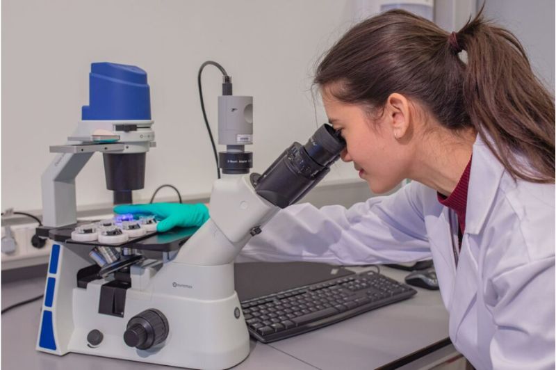
Discover the future of muscle tissue research with the MUSbit™, your gateway to exploring the intricate world of muscle physiology using cutting-edge microfluidic technology.
Ideal for culturing both cardiac and skeletal muscle tissue, this unparalleled microfluidic muscle-on-chip, is a groundbreaking solution that unlocks endless possibilities for researchers.
MUSbit™ is the only microfluidic muscle-on-chip available, which empowers you to advance your studies in unprecedented ways and explore the effects of drug candidates on skeletal diseases thanks to its microfluidic flow channel.
Discover the Power of Microfluidics
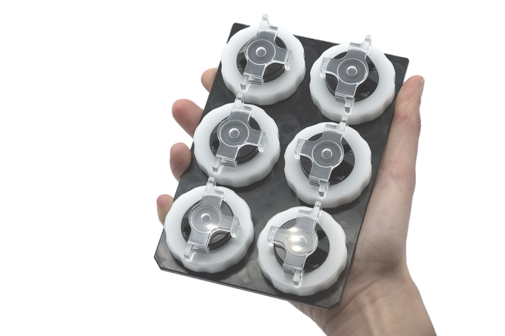
The innovative microfluidic technology of the MUSbit™, combined with our comPLATE™ interface, offers a comprehensive and user-friendly approach to muscle tissue cultivation.
What are the advantages of this unique microfluidic oval-shaped chip:
Versatile Muscle Culturing
The MUSbit™ is designed to accommodate both cardiac and skeletal muscle tissue. This versatile chip is the ideal platform for studying (heart)muscle tissue in a controlled microenvironment.
Precision Engineering
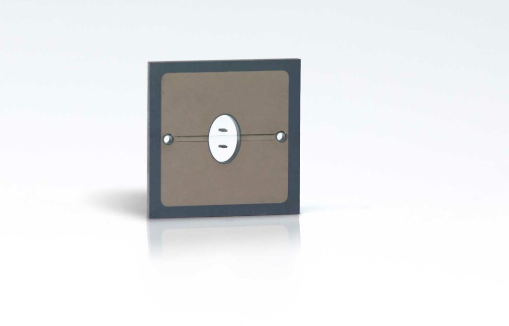
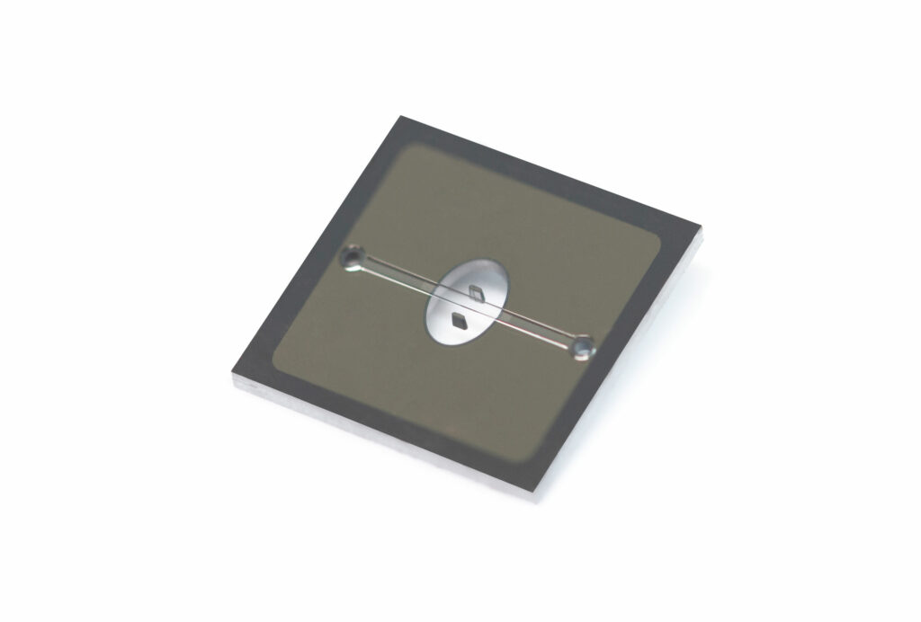
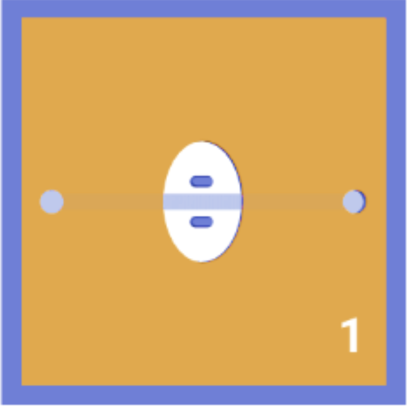
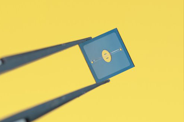
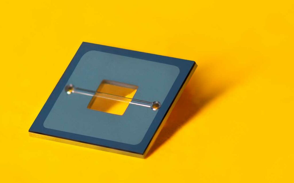
The MUSbit™ features an oval-shaped open well with two pillars intricately linked through a porous membrane. The key to its success lies in the underlying microfluidic flow cell channel, which connects the open well, facilitating precise control over the muscle tissue microenvironment while being able to administer drugs or nutrients to your culture.
Growth Stimulation
The MUSbit™ features an oval-shaped open well with two pillars intricately linked through a porous membrane. The key to its success lies in the underlying microfluidic flow cell channel, which connects the open well, facilitating precise control over the muscle tissue microenvironment while being able to administer drugs or nutrients to your culture.
Continuous Nutrient Supply
The underlying channel within the MUSbit™ serves a dual purpose. It acts as a perfusable blood vessel that continuously provides oxygen and vital nutrients to the muscle tissue, ensuring a thriving microenvironment. Additionally, it is a versatile conduit for administering drug candidates and other substances for controlled experiments.
Optically Transparent Design
The MUSbit™ features an open well that allows direct access to the 3D tissue from the top. This feature, coupled with its optically transparent window on the bottom, enables seamless imaging and real-time monitoring of your experiments.
Testimonials
University of Bari
We choose Bi/ond because we want to develop advanced 3D muscle models for the study of neuromuscular disorders. We believe that this is the best tool to achieve this because thanks to the microfluidic channel on Bi/ond’s chips we can culture 3D engineered muscles and neuron cells in separate compartments. Its compatibility with high-resolution microscopy also allows us to perform our optogenetic assays.
Ornella Capellari
Associate Professor at University of Bari "Aldo Moro"
The Francis Crick Institute
We are using Bi/ond’s platform for the MAGIC” Horizon Europe project we are leading, which aims at accelerating the development of gene therapies and genome editing strategies for muscular dystrophies. The microfluidic channel of Bi/ond’s chips could help us delivering viral vectors while simultaneously perform morphological and functional characterization of the tissue.
Francesco Saverio Tedesco
Senior Group Leader at the The Francis Crick Institute, and Professor at UCL
Unlocking Possibilities with a Microfluidic Flow Plate
The MUSbit™ is not just a microfluidic chip; it’s a complete research solution. When paired with our comPLATE™ interface, you gain access to a safe, user-friendly, and holistic system for muscle tissue cultivation. This powerful combination facilitates experiments that were once unimaginable.
Specifications
The table below shows the dimensions of the MUSbit™ 1 channel. Customization of these dimensions is possible, please contact us for further information.
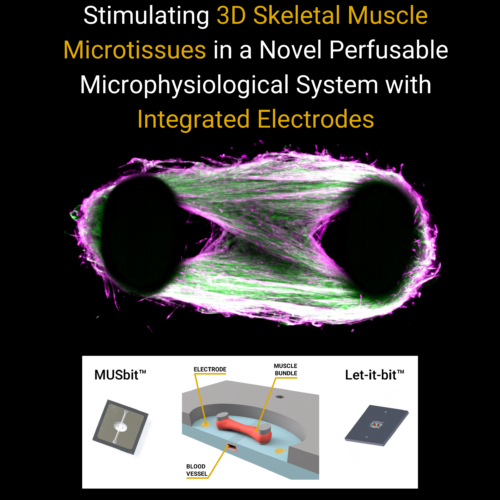
If you’re eager to learn more about our groundbreaking research and how we have successfully cultured 3D skeletal muscle microtissues on the MUSbit™ platform, we invite you to download our informative poster resource. It delves into the details of our perfusable microphysiological system with integrated electrodes, offering valuable insights into our achievements and methods.
The MUSbit™ is not just a tool; it’s a scientific revolution. Join us on the path to redefining muscle tissue research with the unmatched capabilities of double emulsion microfluidics and the MUSbit™ platform.
Bi/ond protects your privacy: when you request a quote, documentation or price options your personal data will be transferred to the Bi/ond team who will be able to contact you directly. See our Privacy Policy for details on the method used to process your data, the purpose and your rights concerning this data. By continuing to use our website you agree to our General Terms of Use.
Bi/ond protects your privacy: when you request a quote, documentation or price options your personal data will be transferred to the Bi/ond team who will be able to contact you directly. See our Privacy Policy for details on the method used to process your data, the purpose and your rights concerning this data. By continuing to use our website you agree to our General Terms of Use.