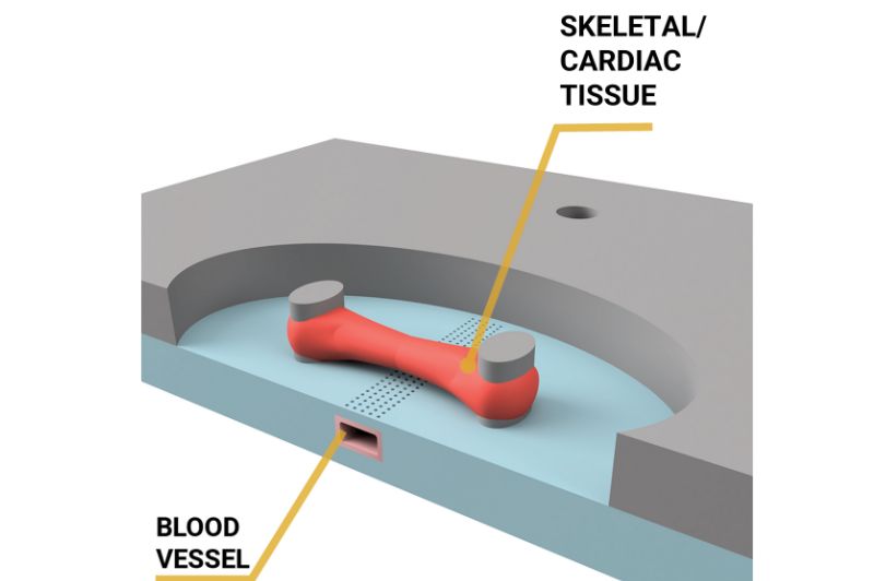
The MUSbit™, in combination with the comPLATE™, presents a revolutionary solution for culturing mature, contractile muscle models of (human) muscle tissue in a controlled and observable environment. This innovative system enables researchers to isolate and study muscle cells, providing a foundation for developing sophisticated muscle models that accurately reflect their physiological properties. Researchers can study the functioning of (heart) muscle tissue, while also testing the efficacy of new drugs, thanks to the microfluidic channel.
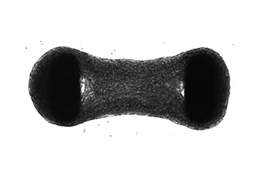
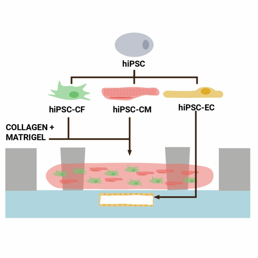
With the innovative open well design, you can pour your cell suspension of muscle cells into the well, watch as they begin to self assemble into a 3D tissue, and witness their contractions in real-time. As the cells start to pull on the two pillars, the shift of these pillars can be monitored to determine the force and rate of contraction.
Nutrient Supply and Drug Administration: A Mimic of the In Vivo Environment
The underlying microfluidic channel integrated beneath the well can be lined with endothelial cells, mimicking the intricacies of a real 3D blood vessel, providing the muscle tissue with continuous access to essential nutrients and oxygen, which in turn facilitates which in turn facilitates that cells self-assemble into a 3D tissue. Moreover, this microfluidic channel also allows for administration of drugs and other substances and subsequent monitoring of any effects on the muscle tissue in the open well in real-time.
Electrical stimulation of your 3D muscle models: developing a heart on a chip
The MUSbit ™ also allows for electrical stimulation of the 3D muscle model, through built-in electrodes, allowing for the recreation of a heartbeat. This remarkable capability provides a powerful tool for studying the impact of various stimuli on muscle function, including electrical stimulation, drugs, and toxins. By mimicking the natural rhythm of contraction and relaxation, the MUSbit™ enables researchers to gain deeper insights into the underlying mechanisms that govern muscle physiology.
Skeletal Muscle on a Chip
The MUSbit™ platform extends its reach beyond the confines of cardiac muscle, venturing into the realm of skeletal muscle research. With the MUSbit™, researchers can successfully culture skeletal muscle cells, generating 3D skeletal muscle microtissues. These complex constructs provide a valuable model for studying the mechanisms of movement, exercise, and muscle repair.
Muscle Models - Applications
With the innovative open well design of our chip, you can pour your cell suspension of muscle cells into the well and watch as they begin to grow in between the two pillars.
The underlying microfluidic channel can be lined with endothelial cells, mimicking a real 3D blood vessel, providing the muscle tissue with continuous access to essential nutrients and oxygen. Moreover, the channel also allows for administration of drugs and other substances and subsequent monitoring of any effects on the muscle tissue in the open well.
Applying external electrodes makes it possible to stimulate the contraction of the muscle bundles in between the two pillars. As the muscle bundles start to pull on the pillars, the shift of them can be monitored to determine the force and rate of contraction. This allows for checking any disruption in muscle function before and after treatments.
Unlike on a 2D model, the 3D model combined with the electrical stimulation, allows for functional characterization and force contraction measurements.
A Collaborative Effort: Towards Breakthroughs in Muscle Research
Our MUSbit™ platform is currently being rigorously tested at the Leiden University Medical Center in the Netherlands, with a publication about the results in the works. Get ahead of the curve and take the first step towards a more comprehensive understanding of (heart)muscle function.
Watch as the black and white video showcases a functional muscle bundle, contracting in between the two pillars of the MUSbit™. Witness the tissue contract natively or in response to cardiac stimulants, suppressants, or even cardiotoxic compounds. The results of this work done in collaboration with LUMC Leiden, Netherlands, are currently under embargo awaiting the world to see its exciting results.
That’s not all, take a look at the stunning fluorescent bundle of cardiac muscles grown between the two pillars of the MUSbit™ on this other video. This bundle has been stained for α-Actin (red, marker for cardiac muscle tissue), cardiac troponin (green, marker for myocardial damage), and DAPI (blue, staining cell nuclei).
Work in collaboration with LUMC Leiden, manuscript in preparation.
Poster downloads
Stimulating 3D Skeletal Muscle Microtissues in a Novel Perfusable Microphysiological System with Integrated Electrodes
Generation of Human iPSC-Derived Duchenne Muscular Dystrophy Skeletal Myocites Suitable for 3D Functional Studies and Investigating methods for Dystrophin Restoration
This poster delves into our successful culture of skeletal muscle cells on the MUSbit™ platform to generate 3D skeletal muscle microtissue. To gain a deeper understanding of our achievements, download this valuable resource now.
In this poster you will discover how researchers created functional 3D skeletal muscle microtissues within the MUSbit, which exhibit twitch and tetanic responses that strengthen over time.
Additional information:
- For more information on our solutions, visit our product and applications pages
- Access our muscle related resources: Poster and on-demand webinar
- To order our products, or to arrange a Proof of Concept study, please contact our sales team.
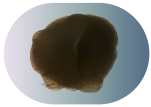
Tumor tissue
Organoids