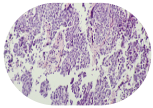
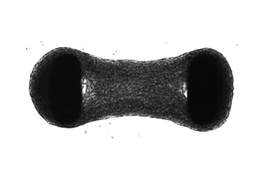
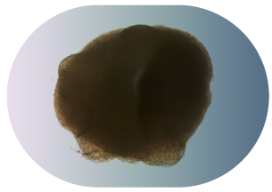
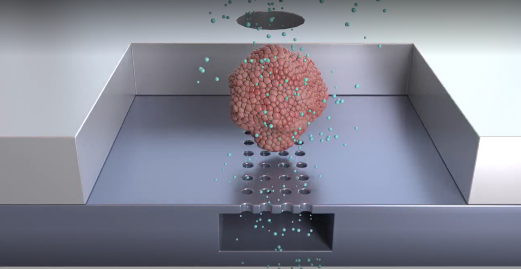
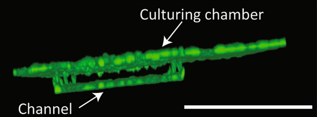
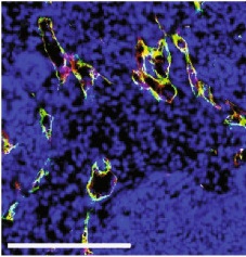
Organoids

Discover the versatility of the Bi/ond organ-on-chip platform with the inCHIPit™ and comPLATE™. Create a human-derived research model that mirrors the organ of your choice like never before.

Organoid image

Experience ease of access to your organoid with the open well of our chip, which allows for direct manipulation from the top and imaging through the clear, optically transparent window on the bottom. The microfluidic channel can be lined with endothelial cells, mimicking a real 3D blood vessel, providing the organoid with constant access to essential oxygen and nutrients. But that’s not all, you can even administer drugs and other substances through the channel.

Get closer to uncovering the mysteries of your research with the Bi/ond organ-on-chip platform!

Organoids and tumor tissue

In practice...

Here you can find an example of how our technology has been used to advance organoid-based research. Our inCHIPit™ platform, in combination with the comPLATE™, has been utilized by leading researchers in Erasmus Medical Center to vascularize kidney organoids and more. To learn more about how our technology has been put into action, you can read the full paper from Bas‑Cristóbal Menéndez et al. 2022 here.

Erasmus MC Logo

Summary:

Kidney organoids derived from human induced pluripotent stem cells (iPSCs) can be a valuable tool to study kidney development and disease, but the lack of vascularization limits long term research.  

In this article, the Bi/ond organ-on-chip system, consisting of the inCHIPit™ 3C with the comPLATE™, was used to culture kidney organoids for 9 days with a continuous flow. Our system supported kidney organoid differentiation and the formation of microvascular structures that fused into perfusable lumens, mimicking normal vascularization.  

Organoids on the Bi/ond platform

GFP+ HUVECs seeded in one of the three microfluidic channels beneath the culture chamber were able to migrate through the porous membrane and formed a monolayer in the culturing chamber, as shown in the picture on the left.

The cells survived 9 days of fluidic flow shearing stress and adopted a directionality in concordance with native endothelium, mimicking vasculature in vitro.


Co-culturing of the organoids in the culture chamber and HUVECs in the microfluidic channel led to the establishment of vascular-like structures with open lumens in the organoid tissue, as shown in the picture on the right. 

This model therefore provides a useful insight into kidney organoid vascularization in vitro and presents a tool for further studies of kidney development and drug testing, both for research purposes and pre-clinical applications.

Organoids fluorescence
“The Bi/ond organ-on-chip platform offers the possibility to culture kidney organoids on top of endothelialised microfluidic channels. This allows us to study sprouting of endothelial cells from the channels into kidney organoids, which represents a first step towards in vitro vascularization of organoids.”
Martin Hoogduin
Dr. Martin Hoogduijn
Associate Professor in the department of Internal Medicine, section Nephrology and Transplantation, at Erasmus Medical Center.

© Bi/ond 2023. All rights reserved.

Bi/ond Logo - Nourishing, Stimulating and Monitoring Cells

Bi/ond protects your privacy: when you request a quote, documentation or price options your personal data will be transferred to the Bi/ond team who will be able to contact you directly. See our Privacy Policy for details on the method used to process your data, the purpose and your rights concerning this data. By continuing to use our website you agree to our General Terms of Use.

Bi/ond Logo - Nourishing, Stimulating and Monitoring Cells

Appplication downloads

Click to download the items.