Tumor Tissue / Cancer Biopsies

The Key to Cancer Research Innovation

In vitro research

Bi/ond’s platform is designed to provide you with a new perspective on cancer biopsies and its implications in the field of cancer research. By harnessing the capabilities of tumor tissue on a chip, you will be able to delve deep into the intricacies of cancer biology.

Transform your cancer research with the innovative inCHIPit™ and comPLATE™ combination. With this powerful duo, you can create a fully tumor-derived model that mimics the tumor microenvironment and get one step closer to unlocking the underlying mechanisms of cancer.

Gain Unprecedented Insights with Optically Transparent Windows

Our platform provides you with direct access to your tumor tissue/cancer biopsy from the top, allowing you to closely monitor the progress of your cancer research. The optically transparent window on the bottom ensures you can image the tumor through them.

Perfusion and 3D Models with Endothelial Cells

With the ability to line the microfluidic channel with endothelial cells, you can create a perfusable 3D blood vessel within your tumor model. This advanced feature stimulates the growth of your tumor, offering an even more accurate representation of the tumor microenvironment. Additionally, it also allows you to test the impact of chemotherapies and other substances on the growth and spread of the cancer. This invaluable tool allows you to conduct precise experiments, shedding light on potential treatment strategies.

A Deeper Understanding of Cancer Complexity

The inCHIPit™ and comPLATE™ are the perfect tools for a comprehensive exploration of the intricacies and complexities of cancer. With these tools at your disposal, you can delve deeper into the complexities of cancer research, gaining insights that were previously beyond reach.

Pioneering cancer research innovation

Our commitment to advancing cancer research innovation is exemplified by our collaboration with the Erasmus Medical Center in Rotterdam, Netherlands. In a groundbreaking study led by Chakrabarty et al. in 2022, we harnessed the potential of inCHIPit™ and comPLATE™ to culture and test breast- and prostate cancer tissue on a chip. The results were nothing short of remarkable, particularly in drug response.

Make sure to check out the full study for detailed insights on how our technology can revolutionize organ on chip cancer research.

Cancer Research

Tumor tissue/Cancer biopsy cultures in organ-on-chip

Cancer treatment faces major challenges in defining the optimal therapy for each individual patient. Although there have been several strategies employed for this, to date, none have been able to reliably predict individual response. Therefore, ex vivo assays that can predict this response would strongly aid selection of the most effective treatment for each patient tumor(s).

In this article, we show how tumor tissue was cultured in the Bi/ond organ-on-chip system, consisting of the inCHIPit™ 3C with the comPLATE™, here referred to as cancer-on-chip (CoC) system. Breast PDX tumor and prostate PDX tumor PC82 slices were either cultured in 6-well standard plates (referred to as ex vivo culture) or in the Bi/ond CoC platform in duplicates.

Scientist removed tissue slices from the chip at either day 7 or day 14 and fixed them in formalin and embedded them in paraffin for microscopic analysis.  First, the condition of the cells in the 6-well ex vivo culture was compared with that of the cells in the CoC platform after 7 and 14 days of culturing.

The obtained proliferation and cell death data indicate that the CoC platform provides better culture conditions for extended culture times  (beyond 7 days) of tumor tissue slices than the ex vivo system. There are already subtle differences between both culture methods even after 7 days, as shown in the figure on the right.

Tumor Graph 7 days

Tumor response to drug treatment

Next, researchers treated breast tumor slices for 3 days with Cisplatin. Tissue cultured in the CoC platform showed a decrease in replicating (s-phase) cells and significant increase in apoptotic cells as compared to ex vivo culturing. In the figure on the right you can see EdU-positive (replicating) cells and TUNEL-positive (apoptotic) cells at day 0 (D0), day 3 (D3) and at day 3 after treatment with Cisplatin (D3Cis). This response to cisplatin treatment correlated well with the known tumor response in vivo as well as in ex vivo cultures.  

A similar response in replicating and apoptotic cells was seen after 7 days of apalutamide exposure of prostate tumor slices. Again, this was comparable to the effect of antiandrogen treatment in in vivo and ex vivo cultures.  

Tumor Drug Response
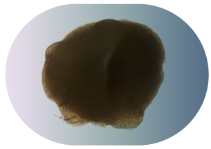
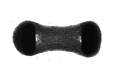
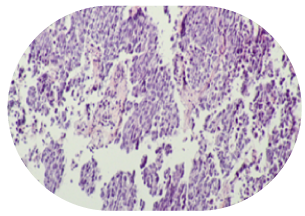
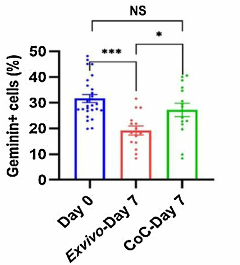
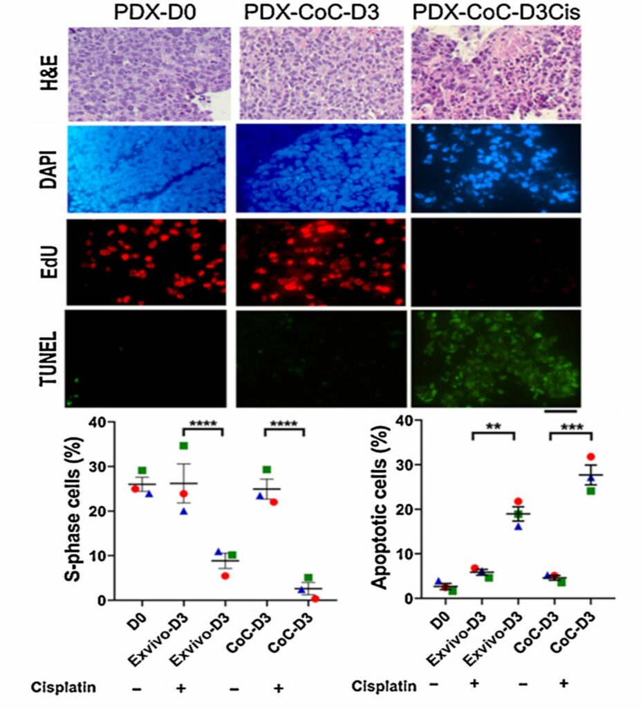
"We are applying Bi/ond devices in therapy response using the breast PDX model. Bi/ond microfluidics chips guarantee accurate controlled condition, which is beneficial for long term tumor tissue slice culture. Setting up the Organ-on-Chip technology went very smoothly in our lab.”
Dijk van Gent
Dr. Dik van Gent
Assistant Professor in the department of Molecular Genetics at Erasmus Medical Center

You can also take critical steps toward a future where we conquer cancer, with the inCHIPit™ and comPLATE™. Contact us today to request our products or get our support for a Proof of Concept

Additional information:

Logo Bi/ond

Bi/ond protects your privacy: when you request a quote, documentation or price options your personal data will be transferred to the Bi/ond team who will be able to contact you directly. See our Privacy Policy for details on the method used to process your data, the purpose and your rights concerning this data. By continuing to use our website you agree to our General Terms of Use.